K

Peptide Research

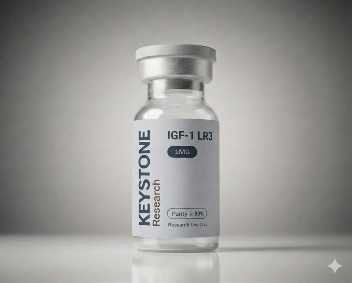
IGF-1 LR3

SKU: IGF-1

IGF-1 LR3 is an 83-amino-acid IGF-1 variant with an arginine substitution at position 3 (replacing glutamic acid) and a 13-amino-acid N-terminal extension. These modifications dramatically reduce binding to IGF-binding proteins (IGFBPs), extending half-life from ~12-15 minutes (n

View Catalog →For Research Use Only
IGF-1 LR3

Mechanism of Action

IGF-1 LR3 is an 83-amino-acid IGF-1 variant with an arginine substitution at position 3 (replacing glutamic acid) and a 13-amino-acid N-terminal extension. These modifications dramatically reduce binding to IGF-binding proteins (IGFBPs), extending half-life from ~12-15 minutes (native IGF-1) to approximately 20-30 hours, and making it roughly 3x more potent. It retains full agonism at the IGF-1 receptor, activating PI3K/Akt/mTOR and MAPK pathways driving protein synthesis and cell proliferation.

Research Studies

10 peer-reviewed studies curated for IGF-1 LR3.

Human Studies2

Human2023PMID: 37261455

Recombinant expression of IGF-1 and LR3 IGF-1 fused with xylanase in Pichia pastoris.

Lu Z, Liu N, Huang H, Wang Y, Tu T, Qin X

Applied microbiology and biotechnology

This research focused on overcoming challenges in producing human IGF-1 and its analog, LR3 IGF-1, which are important for various physiological processes. Scientists employed a Pichia pastoris expression system, fusing both IGF-1 and LR3 IGF-1 with xylanase XynCDBFV to enhance expression levels. Production was scaled up using fermentation in a 15-L bioreactor, and the expressed human proteins were subsequently tested for in vitro bioactivity via cell proliferation assays.

The fusion strategy successfully yielded high expression levels, reaching approximately 0.5 g/L for IGF-1 and 1 g/L for LR3 IGF-1 in the bioreactor. Furthermore, the purified IGF-1 and LR3 IGF-1 demonstrated excellent bioactivity in cell proliferation assays, comparable to standard IGF-1. These results indicate a successful method for producing significant quantities of bioactive human IGF-1 and LR3 IGF-1 for research purposes.

View on PubMed
Human2022PMID: 36091374

Sheep recombinant IGF-1 promotes organ-specific growth in fetal sheep.

Stremming J, White A, Donthi A, Batt DG, Hetrick B, Chang EI

Frontiers in physiology

This study developed and evaluated an ovine-specific recombinant IGF-1 (oIGF-1) to address limitations observed with human recombinant LR3 IGF-1 in fetal sheep studies, such as its low affinity for IGF binding proteins. Researchers conducted in vitro experiments measuring myoblast proliferation and performed in vivo studies using late gestation fetal sheep. The in vivo experiments involved short-term (2 hours) infusion of oIGF-1 to assess anabolic signaling, and a longer-term (1 week) infusion to evaluate its effects on fetal body and organ growth, myoblast proliferation, protein synthesis, and IGFBP expression.

The oIGF-1 successfully stimulated myoblast proliferation in vitro. In fetal sheep, a one-week infusion of oIGF-1 promoted organ-specific growth, notably increasing the size of the heart, kidney, spleen, and adrenal glands, and stimulated skeletal myoblast proliferation compared to saline. Importantly, oIGF-1 infusion did not increase hindlimb muscle mass or muscle fractional synthetic rate, and it maintained similar hepatic and muscular gene expression of IGFBPs one to three, suggesting a targeted growth effect without altering overall IGFBP regulation.

View on PubMed

Animal Studies3

Animal2023PMID: 37114757

Attenuated glucose-stimulated insulin secretion during an acute IGF-1 LR3 infusion into fetal sheep does not persist in isolated islets.

White A, Stremming J, Brown LD, Rozance PJ

Journal of developmental origins of health and disease

This animal study investigated the effects of acute IGF-1 LR3 exposure on insulin secretion. Researchers infused late gestation fetal sheep (n=10) with IGF-1 LR3 or a vehicle control for 90 minutes, measuring basal and glucose-stimulated insulin secretion in vivo. Following the in vivo infusion, fetal islets were isolated (n=6 per group) and tested for in vitro insulin secretion in response to glucose or potassium chloride.

Acute in vivo IGF-1 LR3 infusion significantly decreased fetal plasma insulin concentrations and reduced insulin secretion during a hyperglycemic clamp by 66%. Despite this in vivo suppression, insulin secretion from isolated fetal islets collected immediately afterward was not different from controls. This suggests that while acute IGF-1 LR3 may directly suppress insulin secretion in vivo, fetal beta-cells can retain the ability to recover glucose-stimulated insulin secretion in vitro, with potential implications for long-term treatment modalities for fetal growth restriction.

View on PubMed
Animal2025PMID: 41015370

Revolutionary decellularized Alstroemeria stem-based nerve conduit integrated with GelMA and controlled IGF-1 LR3 release for enhanced rat sciatic nerve regeneration.

Yavuz E, Sağır MS, Ercan A, Erginer M, Barlas FB, Sakarya D

International journal of biological macromolecules

In this animal study, a novel decellularized plant-based nerve conduit was developed from Alstroemeria stem material, integrated with GelMA, and designed for controlled release of IGF-1 LR3. Thirty rats with a 1 cm sciatic nerve defect were divided into six experimental groups for regeneration assessment. Evaluation methods included gait analysis, electrophysiology, histology, and immunohistochemistry, comparing the new conduit to autologous grafts and commercial conduits.

The IGF-1 LR3-controlled releasing decellularized conduit significantly improved axonal regeneration. This conduit demonstrated performance comparable to autologous nerve grafts, without inducing systemic toxicity. These findings highlight the potential of plant-based biomaterials for applications in peripheral nerve repair.

View on PubMed
Animal2019PMID: 31285507

Inhibition of activin-like kinase 4/5 attenuates cancer cachexia associated muscle wasting.

Levolger S, Wiemer EAC, van Vugt JLA, Huisman SA, van Vledder MG, van Damme-van Engel S

Scientific reports

Research explored the efficacy of ALK4/5 receptor blockers SB431542 and GW788388 in preventing muscle wasting, also investigating synergy with IGF-1 LR3. In vitro experiments involved treating C2C12 skeletal muscle cells with vehicle, the blockers, or IGF-1 LR3. For in vivo studies, a C26-CD2F1 cachexia mouse model was used, with mice receiving various treatments including SB431542, IGF-1 LR3, or a combination.

In vitro, IGF-1 LR3, SB431542, and GW788388 all increased differentiation index and mean nuclei count in C2C12 cells. In the mouse model, GW788388 effectively limited the loss of bodyweight, grip-strength, and gastrocnemius weight, and downregulated Atrogin-1 expression. While IGF-1 LR3 treatment also limited muscle mass loss in vivo, this was associated with accelerated tumor growth.

View on PubMed

In Vitro Studies2

In Vitro2012PMID: 22227200

Na+ transport across rumen epithelium of hay-fed sheep is acutely stimulated by the peptide IGF-1 in vitro.

Shen Z, Martens H, Schweigel-Röntgen M

Experimental physiology

This in vitro study investigated the impact of IGF-1 on sodium transport using isolated rumen epithelium from hay-fed sheep and cultured rumen epithelial cells. Experiments measured mucosal-to-serosal and net sodium flux across the isolated epithelium after applying serosal LR3-IGF-1 and examined the effect of amiloride. A separate experiment employed cultured rumen epithelial cells and a fluorescent probe to assess the rate of intracellular pH recovery following acid loading, with and without LR3-IGF-1 and amiloride.

LR3-IGF-1 acutely stimulated mucosal-to-serosal and net sodium flux across the isolated rumen epithelium, increasing net sodium flux by approximately 60% at 20 µg l(-1). This effect was inhibited by amiloride, which, along with observed increases in intracellular pH recovery rates in cultured cells, suggests mediation by Na(+)-H(+) exchange (NHE). These findings indicate IGF-1 is important for the rapid functional adaptation of ruminal sodium transport by increasing NHE activity.

View on PubMed
In Vitro2021PMID: 33938236

Reduced glucose-stimulated insulin secretion following a 1-wk IGF-1 infusion in late gestation fetal sheep is due to an intrinsic islet defect.

White A, Stremming J, Boehmer BH, Chang EI, Jonker SS, Wesolowski SR

American journal of physiology. Endocrinology and metabolism

This research investigated the effects of IGF-1 LR3 on fetal glucose-stimulated insulin secretion in late gestation fetal sheep. Investigators infused fetal sheep (n=8) with IGF-1 LR3 for one week, comparing them to vehicle controls (n=9). Following the infusion, glucose-stimulated insulin secretion was assessed in intact fetuses using a hyperglycemic clamp, and subsequently in isolated fetal islets via static incubations.

Fetal sheep exposed to IGF-1 LR3 exhibited reduced plasma insulin and glucose concentrations, alongside decreased glucose-stimulated insulin secretion in vivo. This impaired insulin secretion persisted in isolated fetal islets, indicating an intrinsic islet defect. These findings suggest that altered insulin/IGF-1 axis signaling may contribute to long-term reductions in β-cell function observed in neonates born with elevated IGF-1 levels.

View on PubMed

Other3

Other2025PMID: 39679943

IGF-1 LR3 does not promote growth in late-gestation growth-restricted fetal sheep.

White A, Stremming J, Wesolowski SR, Al-Juboori SI, Dobrinskikh E, Limesand SW

American journal of physiology. Endocrinology and metabolism

This study investigated the effect of the IGF-1 analog, IGF-1 LR3, on growth in late-gestation growth-restricted fetal sheep. Researchers administered either IGF-1 LR3 or a vehicle directly into the circulation of FGR fetal sheep for one week. During this period, measurements included plasma insulin, glucose, oxygen, amino acids, and glucose-stimulated insulin secretion (GSIS).

Treatment with IGF-1 LR3 did not improve fetal growth, circulating insulin, glucose, oxygen, or GSIS in these FGR fetuses. While insulin concentrations and GSIS were not attenuated, circulating amino acid concentrations significantly decreased in the IGF-1 LR3 group. This decrease in amino acids, particularly branched-chain amino acids important for GSIS and protein accretion, suggests increased utilization and indicates that additional factors may be necessary for IGF-1 LR3 to promote growth in the context of placental insufficiency and fetal growth restriction.

View on PubMed
Other2020PMID: 32573852

Coronary vascular growth matches IGF-1-stimulated cardiac growth in fetal sheep.

Jonker SS, Giraud GD, Chang EI, Elman MR, Louey S

FASEB journal : official publication of the Federation of American Societies for Experimental Biology

Fetal sheep at 127-134 days gestation were studied to determine how IGF-1 LR3 administration affects coronary growth and function alongside IGF-1-stimulated cardiac growth. Animals underwent surgical instrumentation and received either IGF-1 LR3 or vehicle. Coronary growth and function were interrogated using pressure-flow relationships, responses to acute hypoxia with adenosine receptor and nitric oxide synthase blockade, and by modeling coronary flow determinants.

Coronary conductance was preserved on a per-gram basis following IGF-1 treatment, and adenosine and nitric oxide contributed similarly to hypoxia-mediated coronary vasodilation in both IGF-1-treated and Control fetuses. The relationships between coronary flow and blood oxygen contents were also similar across groups. These findings indicate that IGF-1-stimulated fetal myocardial growth is accompanied by appropriate expansion and function of the coronary vasculature.

View on PubMed
Other2021PMID: 33427051

IGF-1 infusion to fetal sheep increases organ growth but not by stimulating nutrient transfer to the fetus.

Stremming J, Heard S, White A, Chang EI, Shaw SC, Wesolowski SR

American journal of physiology. Endocrinology and metabolism

To investigate the role of fetal IGF-1 in placental blood flow, nutrient transfer, and nutrient availability for fetal growth, late gestation fetal sheep (n=8 per group) received a one-week intravenous infusion of either LR3 IGF-1 or saline via catheters. After the infusion period, a metabolic study was conducted to measure uterine and umbilical blood flow, nutrient uptake rates, and fetal protein kinetic rates. This methodology allowed for a direct assessment of IGF-1's impact on various physiological parameters in the developing fetus.

While overall fetal weights were not statistically different, LR3 IGF-1 infusion resulted in higher weights for the fetal heart, adrenal gland, and spleen, along with lower fetal insulin concentrations and increased myoblast proliferation in skeletal muscle. Despite similar uterine and umbilical blood flow, umbilical uptake rates of amino acids and fetal concentrations of multiple amino acids were lower in the LR3 IGF-1 group. These results suggest that LR3 IGF-1 enables the efficient utilization of available nutrients to support organ-specific growth in the fetus, rather than by stimulating increased placental blood flow or nutrient transfer.

View on PubMed

Related Research

Downstream of GH-axis peptides; commonly studied with CJC-1295 + Ipamorelin, standalone Ipamorelin, and Tesamorelin. Paired with Wolverine or GLOW in tissue repair and recovery research protocols.